A group of researchers have developed what they call a "virtual breast" to help clinicians better interpret the results of a cancer detection test - ultrasound elastography.
Like a simulator used to train fledgling surgeons, the virtual breast - a 3D, computer generated phantom - could let medical professionals practice elastography in the safety of the laboratory.
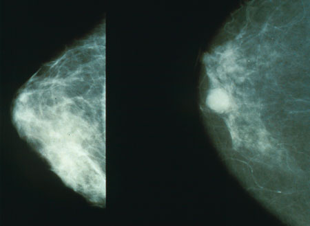
As only a minority of suspicious mammograms actually lead to a cancer diagnosis, the researchers said elastography can be used to pinpoint possible tumours throughout the body, including the breast.
"Ultrasound elastography could be an excellent screening tool for women who have suspicious mammograms, but only if the results are properly interpreted," the study said.
"It uses imaging to measure the stiffness of tissue and cancer tissues are stiff," said Jingfeng Jiang, a biomedical engineer at the Michigan Technological University in the US.
While some of those images can be breath-takingly clear, others are not that precise.
"Depending on who does the reading, the accuracy can vary from 95% to 40%," Jiang added.
As practice could improve better interpretation the results, the researchers developed the virtual breast using data from the Visible Human Project, which gathered thousands of cross-sectional photos from a female cadaver.
It mimics the intricacy of the real thing, incorporating a variety of tissue types and anatomical structures, such as ligaments and milk ducts.
Clinicians can practice looking for cancer by applying ultrasound elastography to the virtual breast and then evaluating the resulting images, the researchers stressed.