The study of conditions affecting the brain is reliant on the fluorescence of the blood circulating within the organ. Induced by dyes, the luminosity helps in investigating the function, or lack of it, in specific regions of the brain for short durations of time. Now, scientists have developed a new method that can help 'light up' blood for several months.
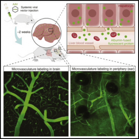
In a new multi-institutional study, researchers have described a technique that utilises a fluorescent protein–a modified form of the blood protein, albumin–that is delivered through a viral vector. Using an animal model, the scientists demonstrated that the vector can trick the liver into producing more of the fluorescence-labelled albumin, causing the secretions to remain in the blood plasma for up to four months or more.
"We developed a new method to visualize the blood flow in the brain in experimental mice for months," said Dr. Hajime Hirase, corresponding author of the study, in a statement. According to the authors, the method can aid in tracking the progression of brain afflictions such as Alzheimer's Disease and strokes. The findings were published in the journal Cell Reports.
Tweaking A Blood Protein

Mice are used in medical research because of the biological similarities they share with humans. They form the basis of several models geared toward the examination of human diseases and conditions, including those affecting the brain such as Alzheimer's disease, Parkinson's Disease, depression, and strokes, among others.
In order to scrutinise the flow of blood in the brains of rodents employed in some disease models, scientists use chemical dyes to induce fluorescence in blood. However, the window for the evaluation of blood circulation within the brain is limited to a few hours due to the requirement of frequent re-administration of these dyes.
Two members of the research team suggested a novel idea to mitigate this constraint. "My graduate students Xiaowen Wang and Christine Delle calculated that if we label a few percent of albumin, we should be able to see green fluorescent blood by microscopy," mentioned Dr. Hirase.
Albumin is the most common circulating protein found in blood plasma. It is synthesized in the liver. Upon entering the bloodstream, albumin aids in the transportation of enzymes, hormones, vitamins, and other important substances throughout the body. It also prevents the leakage of fluids from the bloodstream.
Tricking Liver into Turning Blood Fluorescent

In order to imbue blood with fluorescence, the team attached the gene from a fluorescent protein to the gene in albumin. The altered albumin gene was then introduced into a genetically modified virus that served as a vector. While technically an infectious agent, the redesigned virus does not cause any disease and cannot be transmitted to humans or other animals.
This viral vector containing the modified albumin is injected into mice. Utilising the natural processes of the body, the vector tricks the mice's liver into producing fluorescence-labelled albumin and secreting it into the bloodstream; thereby, making the blood plasma fluorescent.
Highlighting the utility of the new technique, Dr. Antonis Asiminas, co-lead author of the study, explained: "It is a way for us to really study disease in a way that we could not do before. The goal and aim are that it will give us a new insight to disease progression and development in e.g. stroke that can hopefully lead to better understanding and possible treatment." He also added that the method can serve as a tool in analysing the "long term progression of diseases."
Reducing Stress in Animals

Unlike older techniques where multiple doses of dyes are administered frequently to the mice, the new viral vector-based method requires only a single intraperitoneal or intravenous administration. This reduces the discomfort and stress caused to the animals.
"The mouse tail has very thick blood vessels where we normally inject dyes in. Then, if we look in a microscope, we can see nicely labelled blood, but this only lasts for one or two hours. Our new method labels the blood for months," stated Dr. Hirase.
By leveraging a sustainable technique that relies on fluorescent proteins instead of regularly injected dyes, the authors believe that the principles for the ethical use of animals in research, also known as the "3Rs", can be implemented better. The three Rs stand for Reduction (in the number of animals used), Refinement (in testing methods), and Replacement (replacing animals with other testing mediums).
"It addresses two of the three Rs. It's both Refinement, because we are reducing stress for the animals, but it is also Reduction, because we can do longer studies in the same animal repeatedly so reducing the number of animals as well," stated Dr. Asiminas.


